Ever wondered why plants glow after rain? Why rainbows are actually bow shaped? What gives the butterfly its colours or why the stars twinkle? The little moments of 'eureka' that happen in a person's life, changes his perception of things happening around him and leaves him with a desire to explore further. Through this blog we will take you on a journey of thousands of light years into space, explore the invisible world of angstroms, play with atoms and listen to the story that numbers tell.

All narrated in your mother tongue .

हिन्दी मे ... தமிழில்

Showing posts with label BIOLOGY. Show all posts

Thursday, September 16, 2010

On being the largest

I have no idea how it feels to be the largest flower on earth. You will have to ask that to Rafflesia arnoldii. But I definitely know how it smells: like rotting flesh. Yuk! Growing up to 3 ft, there are lots of things that are weird about this plant or should I say flower. For, all that this one seems to have is a flower. No sign whatsoever of stems, leaves (No photosynthesis! Wasn’t it a defining characteristic of being a plant? Got to go back to my 5th std book and make changes) or true roots.


This one belongs to a group of plants called Rafflesia, all of which are parasitic, an endoparasite (grow within the host) to be specific. They specifically parasitize on a member of grape family called Tetrastigma. Rafflesia grow inside the grape plant’s stem penetrating its tissue through a thread like thing called haustorium (Seen in parasitic fungi as well) and absorb all nutrient from it (its like… u make the food, I will put a pipe and suck it out and use it to grow 3ft. Too much of an extravaganza for a parasite).
Once in a while, they put out a bud and after months it flowers, which lasts for few days (~7days). The plant (Rafflesia arnoldii) is unisexual (some species of Rafflesia are bisexual too), so there is a male and female version of the big stinker. Ahh! Now I guess I understand why it stinks! To reproduce it has to pollinate and for that it requires pollinators like flies. And rotting flesh attracts flies. Off comes a fly expecting a feast and Rafflesia sticks its pollens onto its carrier, which unknowingly will transfer the pollen to a female flower. Ingenious! But means by which its seeds are distributed isn’t known for sure, but seems like some shrews eat its fruit (doesn’t that stink?) and thus disperse its seed.

For all this planning and parasitizing, the plant is highly endangered. Blame it on habitat encroachment by humans. That apart there are lots of stuff about Rafflesia that makes them more prone, starting from their requirement of specific host (habitat specificity, occur only in the rainforests of Sumatra and Borneo in the Indonesian Archipelago). Being unisexual, it’s important that the male and female flowers be close by and that they should open around the same time. And to add to it, they flower only for few days, a time constraint as well. What sort of conservation do you think will work out for this flowering giant? (Grow the grape and put some flies and Rafflesia seeds!). Of so many bizarre designs that nature has come up with, Rafflesia arnoldii, is definitely one.

References:
http://www.parasiticplants.siu.edu/Rafflesiaceae/Raff.arn.page.html
http://en.wikipedia.org/wiki/Rafflesia_arnoldii

Monday, May 10, 2010

Dance for navigation

"The bees are buzzin' in the tree, to make some honey just for me…….If you act like that bee acts, uh uh you’re working too hard"- Baloo in the Jungle book

Bees are definitely one of the hardest workers on the planet, with ants being their only close competitors. Bees are social insects, having a hierarchical family group. At the top is the queen and the bottom most are workers who bring in honey to feed the ever growing family. Drones, whose only job is to fertilize the eggs that the queen lays (she actually lays a lot), and larvae in various stages of maturity are the other occupants of the beehive. But why do bees buzz and wiggle around so much, wasting their energy, as if the energy spent on finding and bringing honey wasn’t tiring enough? Well if you have keenly observed bees hard at work, you would realize that all that wiggling and buzzing is their way of communicating with each other. Yes they dance and buzz to convey to their fellow bees the location of a food source In case you never got to observe bees, then let’s do a bit of eavesdropping and find what they do. Make no noise! You don’t want to anger them and get stung.

Get down on the Dance floor


So lets follow the scout bee who is just entering the hive, he seems to be really happy and dancing in a particular way. Well the dance floor is usually near the entrance, but in winters it may move more inside the hive and in real hot climate they use the area outside the hive as dance floor. Since natural hives hang vertically, the dance is also on vertical plane and the bee has to tell a route that’s horizontal, that requires lots of sense of direction and distance (I am already wondering which plane is horizontal and which is vertical). When the dance is outside the hive (or in artificially constructed honey combs), it is generally done on a horizontal plane.

The Round dance

The bee that just entered is making vertical circles. Hey!!! See that he has suddenly reversed direction and is making a circle again. There are so many rapt observers (other bees I mean) besides us.

Let me translate it for you. Circles mean the food is nearby not more then 100 meters away. Having received the good news, the other bees, all fly away in all directions and search within 100 metres radius (hard work), but an experienced bee, using the odour given out by our scout, directly flies to the flower referred to by the scout’s odour (smart work).

The tail-waggle dance

Ohh, we have another arrival! The poor creature looks so tired, but excited nevertheless. So he is taking to the dance floor as well, meaning he too has found food. His moves are different though. Look! He is making a straight run and wagging and buzzing rhythmically (thought to be describing food quality and quantity);turning to left, making a semicircle, back to where he started; he is doing the straight run again, but look, now he is turning right and making a semicircle; making a near 8 figure each time. Some of them have joined in his dance. And the spectators have taken off, so sure of where they are headed. However strange this looks to you, it’s the most ingenious way of conveying the direction and the distance of the food (considering that they can’t speak as we do).
Well, tail waggling dance is performed when food is more than 100 meters away. It uses the angle of sunlight (or partially polarized light*- implies they can convey direction even when the sun isn’t directly visible, say as on a cloudy day or when sun is setting behind a mountain) to describe the direction and number of eights to describe the distance. Bees must be having the most accurate sense of time (since sun and its rays change position as the day progresses; also consider that he has to correct for its position from the time he found food to the time that has lapsed till his dance performance).With increase in distance the number of 8s per unit time decreases and the length and duration increases. So if for a food source 100 meters away the bee makes 10 eights in 15 seconds, for 3km it will make one stately 8 in the same time. Waggle part or the straight run is what conveys the direction. A straight vertical run upwards means the food is towards the direction of sun, downwards implies away from sun. If it is say 30 degrees to sun rays angle, then the run will be at an angle of 30 degrees to the vertical.


Courtesy: www.answersingenesis.org, www.theevidence.org

On days when the dance floor is shifted to the outside the sun itself may be used as reference. Sometimes even the onlookers join in the dance, learning the moves they are actually by-hearting the location.
This language of the bees was first deciphered and translated for us by Karl von Frisch when World War II was on in full swing (when people were decoding other stuff). He understood the bee language so well that he could tell his neighbors the exact distance and location of a flower from which the bees in his side were feasting upon. That’s some marvelous decoding Mr. Frisch! In fact he made artificial honey combs, exposed the bees to artificial polarized light and studied their dances out and out. But that’s not all to it, the dialect and the accent of the bees varies with regions. For e.g. An Austrian bee (von Frisch worked on these) does round dance for a distance up to 100meters, but an Italian bee shifts to tail-waggle after 80meters. So if you made an Italian honeybee talk to an Austrian one, you would most probably end up confusing the Austrian. The bee language is no less complicated than ours but nevertheless well structured to be precise every single time as if a GPS was inside their head.
* Tie a rope to a pole at one end, stretch it to its length and pluck it. It forms ripples in only one direction, the direction along which it was plucked. Similarly polarized light waves also vibrate only in one direction (light waves are polarized by polarizer and also by scattering). Sunrays get scattered by molecules in atmosphere and become partially polarized. Although the amount is going to vary with region and the sun’s location in the sky, the pattern is very much predictable. And it’s these patterns that the bees recognize and use for their communication.

Reference:

Bionics,Vincent Marteka;Lippincot, 1965

Sunday, April 18, 2010

μίτος (Mitos)

Every one of us starts our life’s journey as a single cell. How do we grow up to become what we are? (Of course several other processes go into making a human being but just consider the sheer increase in the number of cells in your body from 1 to billions). You fall down playing football injure your knee, but after few days the knee looks as it was before. How does mould grow on your bread to such huge amount when you had left it just for few days? Let’s do a bit of time traveling and try finding out.

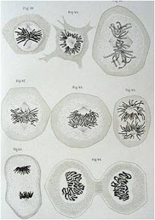
It’s late 1870s (somewhere around 1876-77). Enter the University of Kiel, Germany; into the lab of Prof. Walther Flemming. He pioneered the use of aniline dyes to stain and observe the nucleus. His discovery – the chromosomes (Greek for colored bodies) would become one of the 10 most important discoveries in cell biology. He seems to be busy observing something under the microscope and making some drawings. Come closer let’s have a look at what he is up to. Can you see the red colored thread (figure below are sketches made by Prof. Flemming, with black ink though) like structures at different locations in different cells? They are nothing but the chromosomes, the packaged form of DNA. Walther Flemming is observing and recording various stages of cell division for the 1st time.
Courtesy: Wikipedia

What he had seen in the 17th century is mitosis (From Greek word Mitos- for thread). Mitosis is the process of multiplying a cell’s chromosome into two sets which is generally followed by division of the cell into two daughter cells (cytokinesis), each one getting one set of the chromosome.
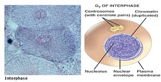
To embark on the journey of mitosis (cell division) the cell has to equip itself. This is what it does in the interphase (or the in between phase-in between two mitosis). The entire cell’s machinery starts to work in full swing. Lets have a 1st hand information from the cell himself as to what he is upto.

“Hello there! Oh don’t get intimidated by the hurry burry happening around. Lots of work going on…. got to prepare for the big event you see. So I am taking in the nutrients, making proteins and lots and lots of energy, soon I will have to start duplicating my DNA. By the way, I am currently in G1 phase of interphase (that’s what you humans call it). Sometimes some of my brother cells decide to take a break at this phase (may be because of lack of nourishment or bad conditions around) and do not go ahead to synthesis DNA. But no breaks for me now, so I am going ahead with DNA synthesis such that there are two copies of my DNA (S phase). I got to distribute the DNA equally to my daughters. That’s a tiring process, takes up huge energy to do that, since I have to ensure no mistakes are made else my progeny will pay heavily for it. But since everything till now has gone well I can’t afford to rest now, have to keep synthesizing proteins (G2 phase) that will help in the mitosis. For successful occurrence of interphase and mitosis (that is yet to occur), the credits go to two groups of proteins that are mainly responsible for it, which together are called the cyclin-CDK complexes.
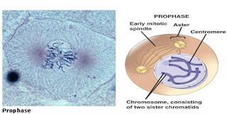
Now I am all set to start off mitosis. The 1st step in the process of mitosis is the prophase (the before stage). As you might have already noticed, the DNA, which was present as loose coil (chromatin) till now, has started to condense, forming the chromosome (dividing/distributing is easier when things are compact). The two copies of each region of the chromosomes also appear in doublets (sister chromatids) attached at a region called centromere. The architect who helps maintains shape and structure - the centrosome - jumps into action (centrioles making up the centrosome have also replicated during the interphase) and you can see the centrioles going to the two poles of the nucleus, as things proceed centrioles are going to be doing the chromosome separating job.Whoosh goes the nuclear membrane!!! I have broken it down so that for the 1st time ever, in my existence, the nuclear contents are going to mingle with the rest of the regions that make me up. It is now prometaphase. Can you see centrioles and the chromosome playing search and capture game? Well that’s actually a serious business. Some of the microtubules (now also called the mitotic spindles) bind (with the help of motor proteins) at the centromere (kinetochore microtubules), few at the arms of the sister chromatids (astral microtubules) and still others with the spindles arising from the opposite poles (nonkinetochore microtubules).
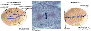
Into the metaphase, which is exactly half way through the process you can notice that my chromosomes are getting aligned one by one along my centre plane. They are all set to be separated into two, once the check proteins finish their last minute testing to ensure everything has been fine till now (similar to the checking before a rocket launch that you people do). As I had told you before, I cannot afford any mistakes. The final GO is yet to arrive from the kinetochore (which is the centromere bound by various proteins). Ahh!! There…. got it.

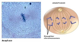
Now my next task is to separate out the sister chromatids (named Anaphase, Ana in Greek means up. Weird how humans come up with such names!) and bring them to the two poles. Any guesses how I am going to manage that? Well I have got a simple old strategy which I learnt from my mom. Make one set of the microtubules, which is holding the chromosomes at the centre, short and the others to elongate. Yes creating a pull and a push force, ripping the sister chromatids to two (Its now daughter chromatids) and stretching the cell as well. I will be handing down each of the pair of chromatids to my daughter cells.
Oh boy! Oh boy! I can’t contain my excitement. I have finally reached the final leg (Telophase) of my journey and all set to give the finishing touch to this long and tedious but well planned and executed journey of mine by first forming a nuclear membrane around the two segregated set of chromosomes.”
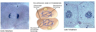
Well what happened next to our cell is anybody’s guess. He distributed his contents equally to two daughter cells in a process called cytokinesis (considered to be separate from that of the phases making the process of mitosis). A kind of constriction (cleavage furrow) occurred around the region where the chromosomes were present during the metaphase, to bring about the separation. (Cytokinesis usually starts off along with telophase and by the time the nuclear membrane forms completely the cell has also divided into two with all the organelles and the constituents in a parent cell, divided equally among the daughter cell).

There you go, the cell just showed you a way it has learnt and perfected through the course of evolution to increase in number, a means of replacing the dying cells, a process employed in making you (development of all multicellular organisms) and repairing you.

Reference :
Molecular biology of the cell. Bruce Alberts, Alexander Johnson, Julian Lewis, Martin Raff, Keith Roberts, and Peter Walter. Garland science 4th edition.

Wednesday, March 3, 2010

The Cell City

Welcome to the ancient city called the eukaryotic cell. Through this guided tour I will take you to various tourist spots and give you an overview of what role they play in sustaining this city. Although ancient, it is an ever evolving city equipped with all modernity. All the major places in the cell are in general called the organelles. Let us now enter the city.

THE CELL (modified from enchantedlearning.com): A Roadmap

Cell membrane :The City wall. The city is surrounded on all sides by this highly guarded wall. At the gates are the watch towers, called the channels, made of proteins which check the identity of every molecule entering and exiting the cell. And not everything can pass through it. The cell membrane is mainly made up of lipids and proteins. Some small molecules though can easily pass through the membrane especially if they are small lipid molecules, similar to the ones that constitute the membrane. Plant cells, fungi and certain bacteria are further protected by another stronger structure called the cell wall. Unlike cell membrane they are made of cellulose. Let me zap you to 3nm size and make you lipophilic(the lipid molecules will like you this way and allow you enter the city)

Cytoplasm: Stick together and keep up with me, now that we are entering the hustling and bustling streets of the cell. This is called the cytoplasm. And all those molecules (ATPs, glucose, oligosaccarides, amino acids, RNAs) are rushing around to carry out various activities required for sustaining the cell. Some of them simply diffuse across and other more important ones take the cell’s railroad called the microtubules and the actins.

Nucleus: This is the administrative centre of the cell, where all the major decisions for running the city smoothly are made. Designing, planning and execution of all the laws and strategies are done in accordance to the constitution called the DNA, which contains the complete information to run any given cell city. Only some of the rules are used to run a given city (called the cell type). The nucleus is also surrounded by a wall called nuclear membrane and heavily guarded. The sign post reads RESTRICTED ENTRY. Only the things that the nucleus requires or asks for can enter into it. The sentries here are proteins called porins who check for special ID (tags) on each of the molecules awaiting an entrance. The information in the rule book to be conveyed to the rest of the cell has to be converted to RNA (a language understood by other components in cell) which is then translated to proteins, which are the actual executioners of the information conveyed by the DNA. Apart from DNA, nucleus has one more component called the nucleolus.

Nucleolus: The assembly of the components of an important machine of the cell, the Ribosome, happens here. Why so much care and importance for just this one organelle? Maybe because ribosome is one of the most important machines in the city, which helps in making proteins.

Ribosome: Now, have a look at one of the finest and efficient machine you will ever see- the ribosome. These are responsible for the production of proteins from amino acids (the building blocks of proteins). The mRNAs (messenger RNA, which carry the message/information from DNA) come here and the Ribosome recruits RNA called transfer RNAs (tRNA) which bring in the relevant amino acids to help manufacture proteins (translating the message from DNA). The platform for ribosome to manufacture its products is provided by the endoplasmic reticulum(ER).

Endoplasmic Reticulum (ER): The industrial park. Here is where the protein-making ribosomes are also located. ER is a chain of industries involved in processing and storage of proteins. From the ribosome the proteins move into the ER, where they are helped to assume their final structure (structure of the protein is essential for its function) and some proteins get processed further. During processing various changes like glycosylation (attaching sugar units to protein) is carried out, that are required for the proteins to do their respective jobs. Of course only proteins with tags and the glucose units are allowed inside. Structurally ER is a huge membranous network and hence the name. There are two types of ER: smooth ER and rough ER. Rough ER is so called because it is studded with ribosome on its surface and hence mainly work as processing units and smooth ER (without ribosome) is mainly for storing proteins. Some cells of your body like the muscle cells have a special type of ER called the sarcoplasmic reticulum which acts as reservoir for calcium ion.

Mitochondria: The PowerStation. The functioning of the city requires power supply which is manufactured here inside the mitochondria. Mitochondria have more roles to play in the cell apart from energy generation. You can read more on it here. Some cities like the cells of plants have an additional solar power plant called the chloroplast that uses the sunlight to produce energy (ATP and NADPH) and which in turn is used for producing sugars.


Golgi apparatus: The Postal service. Golgi bodies can process lipids and proteins from ER and sort them to various regions (called “vesicular transport” in case of proteins from ER). Like the post offices, golgi adds address (labels) to the proteins and lipids which are then transported by the cells transportation system comprised of microtubules and actins to their destinations on time.

Lysosomes: The recycling plant. The cell is maintained spic and span by these. They eat up the old organelles, the bacteria that are engulfed by the cell and other cell debris. Lysosomes are loaded with acids(pH 4.5, cell’s pH is 7.5) and enzymes which act as cleaners. Apart from cleaning, they can also help the cell in digesting the ingested food (similar to what your stomach does). There is one more type of recycling plant called the peroxisomes which are very similar to lysosomes. These are the detoxifying units where the toxic peroxides are got rid of.

Centrosomes: Meet the architects of the city. Also called the microtubule organizing centre, it is the region from which microtubules are manufactured for connecting the various regions of the cell. Centrosomes not only help in connecting the regions of cell through the railroads but also look after the maintenance of the structure of the cell. They are also the junction where all railroads converge. Apart from this, centrosomes are vital during cell division (mitosis and meiosis). They form string like structures called mitotic spindles that help in pulling apart and hence distributing the chromosomes (Condensed DNA) equally to the daughter cells.

Vacuoles: Lastly the warehouses, where the food particles are stored. Even engulfed bacteria to be later thrown out are kept segregated here. They also serve as water towers and also play a role in exporting things outside the cell. Their exact usage depends on the type of cell concerned.

That’s it folks, a small tour around the cell city. Hope you enjoyed it. On exiting through the cell membrane you will be zapped back to your original size. Looking forward to meeting you again.


Friday, February 5, 2010

Bubbling with energy

Millions of years ago when the life was still unicellular and eukaryotic cells had just started evolving, one eukaryotic cell had a prokaryotic cell for its desserts. Lo and behold! the prokaryotic cell could carry out respiration for the predator, producing large amounts of energy; amounts that the predator had never seen before (anaerobic glycolysis was the only route of energy production till then). So the predator cell established a symbiotic association with its prey and in the course of evolution the prokaryotic cell has become a part of all the cells and has come to be known as the Mitochondrion. This widely accepted hypothesis of mitochondrial origin is called the endosymbiotic theory (All circumstantial evidences support this theory, though there is no direct experimental proof). Other theories like the autogenic origin claims that mitochondria was slowly evolved by the eukaryotic cells themselves like other organelles to have more organized and efficient energy production or that it arose from endoplasmic reticulum(ER) and slowly acquired its components. However had been the way mitochondria came into existence, it has become a vital organelle for the very survival of any eukaryotic cell, not only producing energy, but also participating in an array of functions that happens in a cell.

Appearance:

Mitochondria come in many shapes- oblong, branched, oval or circular; the shape varies with cell type and its health. But all of them have two membranes encircling them- the outer membrane and the inner membrane. Outer membrane allows free movement of molecules from cytoplasm in and out of it. The inner side of inner membrane is the highly folded (like those wrinkles of your grandpa) cristae. These wrinkles provide a large surface area tucked into the small organelle (0.5-10 µm), since this is the site of almost all actions happening inside the cell’s energy generator. The inner membrane and the space enclosed by it (the matrix) contain all the proteins, and molecules (O2 being one of it) to carry out respiration.

Interior view of the mitochondria- Top: electron micrograph of mitochondria, down: cartoon depiction of mitochondria.

Division:

Housed inside the matrix is the mitochondrion’s DNA which can help it make some of the enzymes that it requires to carry out the energy production (rest of the information is embedded in DNA in the nucleus). Mitochondrial DNA undergoes replication (make a photocopy of itself) .and like a bacteria or yeast, a mitochondrion can undergo fission to form two mitochondria (with DNA distributed equally between them). If a mitochondrion grows old and loses its DNA it is eaten up by cell’s cleaning system called lysosomes (similar to phagocytes which are the scavenger cells in the body). In a given cell the number of mitochondria is maintained constant; not just by fission, but also by fusion where two or more mitochondria come together and form a new mitochondria.


Job profile:

Powering the cell:
The most important thing that the mitochondria do is to produce energy. The food that you eat is broken down in your digestive system to glucose and transported to the cells of your body. This glucose is used by the cells to produce energy for all its work. The energy in the cell is in the form of ATP (Adenosine triphosphate). Prokaryotic organisms like bacteria which lack mitochondria also use glucose and produce energy, so why have mitochondria in the first place? Mitochondria can help in producing more number of ATPs from the same number of glucose. One molecule of glucose can be used to make ~30 ATP molecules by the mitochondria (cells without mitochondria can make only 2 ATPs for every glucose molecule used)
Mitochondria are very similar to the power plants generating electricity. Glucose is like the coal used to boil water into steam; here glucose transfers its energy to NADH (a coenzyme involved in redox reactions) through a series of reactions called the glycolysis and the Krebs’ cycle. NADH in turn transfers its energy ( in the form of electrons) to the molecules forming the electron transport chain with O2 as the final electron acceptor . In the process a proton pool on the outer side of the inner membrane is generated. These protons (like the steam driving a turbine), drive ATP synthase (consider it as a fusion of turbine and generator), to synthesis ATP from ADP.

Calcium buffering:
Ca2+ is a very important signaling molecule for the cell, levels of which regulate an array of reactions happening inside the cell. Hence keeping a balance of its levels inside the cell is very important. Too much of Ca2+ roaming around freely in the cell can even kill a cell!!! Mitochondria like the other cell organelle endoplasmic reticulum can store Ca2+ within them temporarily when not required and release it as and when the cell asks for it.

Death signal:
For the good of the whole, sometimes a few cells which have grown old or are defective have to undergo death by suicide called apoptosis. When a cell sense (pro-apoptotic stimuli) something wrong with itself it embarks on a series of events that finally will lead to its death. Mitochondria play a significant role in sensing and participating in these changes. Mitochondrial inner membrane contains a molecule called cytochrome C (Cyt C). When mitochondria gets green signal for death, Cyt C is released from it. Cyt C then activates enzymes( caspases) which help in breaking down the cell starting from its DNA to other organelles packaging each of them into apoptotic bodies , which are neatly packed cell debris and are cleared by phagocytes.

To carry out all these functions, the mitochondria moves around in the cell wherever they are needed. Consider a protozoa swimming towards its prey, it requires more energy in the region where it is throwing its pseudopodia. So, more number of mitochondria will be taken to that area by the microtubules , so that there is no shortage or delay in the supply of ATPs. Further, new ones are brought to the scene of action, when the previously present mitochondria gets exhausted (these are moved back into the cell again where they are given time to rejenuvate). This double membrane organelle, however its origin might have been, is an indispensible character in the cell, keeping the cell healthy and bubbling with energy.

Friday, January 1, 2010

Viruses, Viruses and Viruses!


“Mannu don’t play in the rain, you will catch cold” calls out Mannu’s mother as he gets drenched in rain. During the rainy season a number of children get diagnosed of ‘Common cold’. Similarly, there is an increase in the number of ‘Chicken pox’ cases during the summer season. The recent outbreak of Swine flu caused havoc throughout the world. All these are human diseases caused by viruses: common cold is caused by Rhinovirus or Coronavirus, chicken pox by Varicella-Zoster virus and swine flu by H1N1 virus. It is known that viruses are unable to grow and reproduce outside a host i.e. they are non living outside the body of a living organism, then how can they spread and survive in the atmosphere? Scientists, till date, debate whether viruses are living or non living. Such a simple question could not be answered yet, as it raises a fundamental question - what defines life?


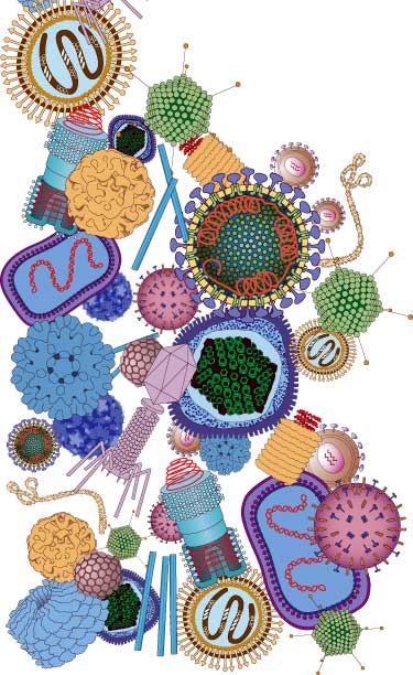
So before we get on with finding out the answer to that simple question, let us see what these viruses are made of. ‘Virus’ is Latin word for ‘Poison’. They are well established as parasites of living beings. They are much smaller than even bacteria and their size ranges from 20nm to 300nm (1nm = 10-9m; cannot be seen using a microscope!). A virus simply consists of a core of nucleic acid (DNA or RNA) enclosed within a protein coat called Capsid. Some viruses have an extra covering made of lipid membrane called Envelope (e.g. HIV- which causes AIDS), but they do not have any machinery for carrying out metabolic activities. Even the simplest and most primitive unicellular organism, the Blue Green algae (also known as Cyanobacteria, 2.8 billion years old fossil samples have been found!), contain protoplasm with membrane bound organelles participating in energy consuming processes. Viruses replicate themselves by utilizing the host cell machinery in a sequence of stages (consisting of adsorption, penetration, uncoating, nucleic acid replication, maturation and release). During Adsorption , the whole virus lands and fixes itself on to the surface of the host cell. This is followed by Penetration in which the Nucleic acid of virus alone enters in to the host cell leaving the protein coat behind. Once inside the cell, the viral genome (nucleic acid) integrates itself with the host cell genome and as the host carries out its normal round of replication, unaware of the presence of viral genome, replicates the viral genes too. These viral genes which code for viral proteins get expressed and result in the formation of many new virus particles. The viruses taken together have genes to counter attack every known form of immune response against them by the host. This is how a virus propagates itself. While outside a host cell it remains dormant waiting to come in to contact with living cell. Air borne viruses like the cold virus remain alive only up to 3 hours outside human body, after which they cannot replicate themselves even when in contact with host.


A virus seems to be on the verge of life, similar to a seed i.e. it looks dead but possesses a certain potential for life. Viruses also have been known to constantly evolve themselves as seen by emergence of new viruses like Swine flu etc. The Eighth report of the International Committee on Taxonomy of Viruses (Taxonomy is the science of classification) reports existence of more than 5450 viruses belonging to more than 2000 species. These viruses infect all type of cells, ranging from bacteria (they are called Bacteriophages) to blue green algae (Cyanophages) and also human beings. A recent report shows that marine viruses help in increasing efficiency of photosynthesis in blue green algae, contributing up to 5% of total world’s oxygen. Apart from this various other genes of viral origin are identified in human beings too, indicating that viruses play a major role in evolution of life on earth. Thus in a way they control life on earth by determining the survival of their hosts. These so called non living beings are closely intertwined in the web of life making them immensely powerful.

Coursing through the Cell

Peeking into a cell using an electron microscope you can see many compartments which are called organelles. Each of these organelles is a mini factory producing goods required for the happy functioning of the cell. Each one of them manufactures unique products (proteins, RNAs, sugars etc) that are distributed to others either to be utilized or to be processed further. To ensure safe and timely transport of these precious consumables, the cell uses a very intricate rail system consisting of five specially designed protein molecules, two of which, called actin filament and microtubule, act as the railroads, and the rest three as the goods carriers.

Like the local trains connecting places across the city, all the regions in the cell are interconnected by actin filaments which are randomly distributed throughout the cell. The cargo in this network is transported by the motor protein called myosin (one of the goods carriers) to their respective destinations. This line connects even places where the second type of rail road, the microtubules, is unable to access. Microtubules are like the interstate expresses connecting only selected places and unlike actin, microtubules are more neatly organized. Kinesins and dyneins use this route for delivering the cargo. Kinesin mediated transport generally is used to bring cargo to cell’s periphery and dyneins ,towards the cell’s interior. All of these motors are powered by ATP (adenosine triphosphate, an energy rich molecule produced by the cells).

Each of these motors, although different in structural and functional details, share some common features. They all have heads and a tail connected by a central region called stalk. They carry their goods by their tails and hold on to the rail (actin filament or microtubules as the case may be) using their heads. On their heads is also the region where ATP can bind and gets hydrolyzed to give the energy for moving the motor.


Myosin keeps a 10nm step for every single ATP used (nearly 3.048 x 10⁻⁹ times your step size), Kinesin and dynein have a 8nm step size.(remember the late Michael Jackson doing his famous moon walk , nearly that’s how the motors look when they move along the rail). Check out these videos to watch myosin and kinesin on the move:


Among the three goods carriers, myosin and kinesin give better performance than dynein . The amount of ATP in the cell affects the working efficiency of dynein, whereas kinesin and myosin are independent of this factor. Moreover dynein’s step size also varies with the load its carrying and availability of ATP. Dynein also tends to take backsteps even in the absence of a load. But it overcomes these drawbacks and comes in par with myosin and kinesin by making use of additional proteins such as dynactin which are like the additional engines that provide the extra force and support to a goods carrier travelling in hilly regions.

It has been observed that all of these carriers work together and sometimes also help each other carry the heavy load, especially dyneins almost always teams up and work together. Given that both the railroads are such a busy network, how is the traffic on the lines regulated? It may not be such a great trouble on actin filament line as it’s only the myosin using the route and also these lines are more numerous. But when it comes to microtubule, two different goods carrier (kinesin and dynein) use the same line! To add to the problem these two carriers move in different directions. Although not much is known as to how traffic jams on a single line are avoided, it is clear that dyneins can avoid traffic by changing tracks.If a kinesin and a dynein happen to take the same line, kinesin being a lean and mean machine forces dynein to change its track.

All this said about the cell’s railway system, three important questions remain unanswered as yet; how does the goods carrier know its destination? And how are head on collisions prevented (Accidents don’t occur in healthy cells)? While functioning together how do they co-ordinate with each other (ensuring that the cargo doesn’t get lost and that ATP doesn’t get wasted)? With the amount of active research going on in this field we can hope to have the complete understanding of the cell’s railway system soon.